Structural accommodation of K at dravitic tourmaline’s X-site: insight from Raman spectroscopy and single crystal X-ray diffraction
Previously Published Material: The Raman spectra were presented at the IMA conference in Johannesberg, September 2014.
Abstract ID#: 36068
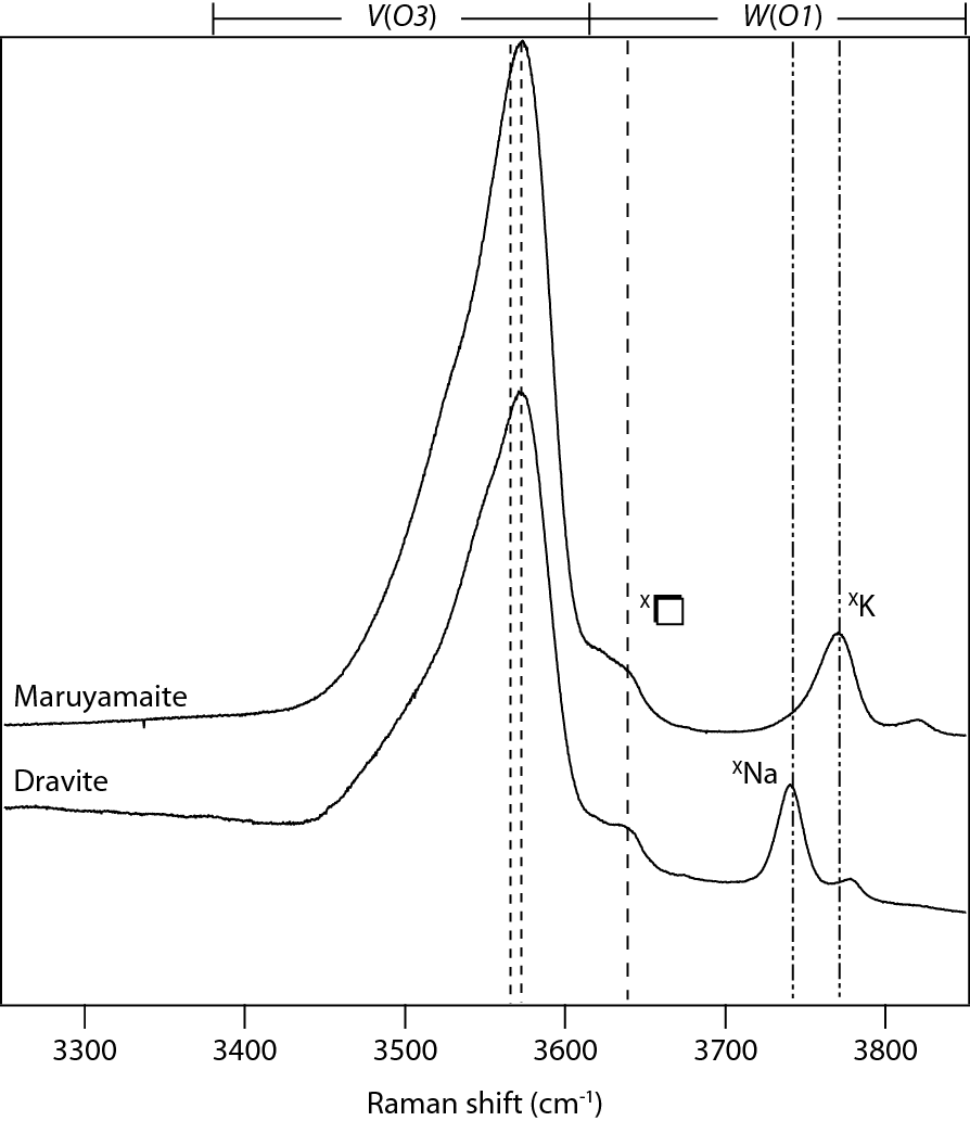
To understand how tourmaline accommodates the K+ ion, we characterized synthetic maruyamaite by electron microprobe, single crystal and powder XRD, Raman and IR absorption spectroscopy. Our synthetic tourmalines have a constrained composition and correspondingly simpler Raman spectra allowing us to unambiguously assign the O-H stretching bands to specific site compositions. The incorporation of K in dravitic tourmaline expands the unit cell volume from a value of 1570 Å3 for our dravite to 1588 Å3 for our maruyamaite. In detail, the X-site polyhedra of the latter expand, with average <X-O> distances of 2.737(12) Å (cf. ~2.69 Å for dravite). The other coordination polyhedra are similar in size to dravite. Comparison of the Raman spectra of dravite and maruyamaite reveals K’s effect on the local bonding environment. The Raman band assigned to the occupied X-site shifts from 3750 (dravite; XNa) to 3769 cm-1 (maruyamaite; XK), reflecting the shortening of the O-H bond in the X-site-neighbouring W(O1) site. In addition, the presence of vacancies at the X-site is common to all synthesized tourmaline, as indicated by a band at 3618 – 3622 cm-1. The lower frequency of this band indicates that vacancies are associated with a lengthening of the O-H bond at the W(O1) site.
The investigation of Raman spectra of well-characterized synthetic tourmaline allows unambiguous band assignment. Moreover, the occurrence and relative positions of these bands combined with single crystal XRD provides insightful information about the effect of composition on tourmaline’s crystal structure.